
جراحی های استخوان و اصلاحات ساختاری |
Bone surgeries and structural corrections

پیوند استخوان و لیفت سینوس
بطور كلي در بيماراني كه بدليل بيماري پريودنتال پيشرفته دچار تخريب استخوان آلوئولار شده اند در جراحي هاي بازسازي يا رژنراتيو پريودنتال، پيوند استخوان بر اساس ميزان و شكل تخريب استخواني انجام ميشود كه به آن GTR گفته ميشود. پيوند استخوان ميتواند با استفاده از استخوان خود بيمار انجام شود و يا از مواد جايگزين استخوان به اين منظور استفاده شود. رايج ترين مواد جايگزين بشكل پودر استخوان در سايز ذرات بين ٢٠٠٠-١٥٠ ميكرون هستند. علاوه بر پيوند استخوان، استفاده از غشاي سد كننده براي مهر و موم كردن ناحيه پيوند از اصول درمانهاي بازسازي استخواني است.
در بيماراني كه كانديداي درمان ايمپلنت دنداني هستند ولي استخوان فك در اثر زمان طولاني بي دنداني، سابقه ي بيماري پريودنتال، يا عفونت در ناحيه ي دندان كشيده شده ضعيف باشد، پيوند استخوان ضرورت پيدا ميكند كه به آن GBR گفته ميشود.پيوند استخوان فك تحليل رفته به منظور كاشت ايمپلنت ممكن در بعد عمودي، افقي، يا هر دو لازم باشد. يعني ارتفاع، عرض، يا هر دو دچار تحليل باشند. در ناحيه ي دندانهاي مولر و پرمولر فك بالا يكي از محدوديتهايي كه وجود دارد ميزان گسترش فضاي سسنوس به سمت پايين يا به اصطلاح پنوماتيزاسيون سينوس است. در بي دنداني هاي طولاني، سينوس ماكزيلا كه حفره اي تو خالي است با ديواره هاي استخواني و پوشيده شده با يك غشاي مخاطي ويژه بنام غشاي اشنايدر، شروع به بزرگ شدن ميكند. كف سينوس ناحيه اي است كه بايد فاصله ي كافي (حداقل ١٠ ميليمتر ) از لبه ي استخوان فك داشته باشد در غير اين صورت نميتوان ايمپلنتي با طول استاندارد قرار داد. جراحي پيوند استخوان كف سينوس كه اصطلاحا به آن جراحي sinus lift يا sinus augmentaion surgery گفته ميشود روشی است که در آن با ايجاد يك حفره ي دسترسي از ديواره لترال يا بيروني سينوس يا از طريق كرست استخوان، بدون آسيب زدن به غشاي اشنايدر، پودر استخوان به كف سينوس پيوند شده و كاهش عرض و ارتفاع استخوان براي كاشت ايمپلنت جبران ميشود.
يكي ديگر از موارد مهم انجام پيوند كه امروزه بسيار رايج است پيوند استخوان در دنداني است كه كشيده ميشود و قرار است طي دو تا چهار ماه بعد كاشت ايمپلنت انجام شود. در اين موارد به كمك جراحي socket preservation كه شامل پيوند استخوان و قرار دادن غشاي سد كننده است، استخوان كافي در حفره دندان كشيده شده تشكيل ميشود و متعاقبا كاشت ايمپلنت با تاخير كوتاهي انجام ميگيرد .

Bone Grafting and Sinus Lift surgery
In general, for patients who have suffered alveolar bone loss due to advanced periodontal disease, bone grafting is performed during periodontal regenerative or reconstructive surgeries based on the extent and pattern of bone loss. This procedure is referred to as Guided Tissue Regeneration (GTR). Bone grafting can be done using the patient’s own bone ( autogenous graft), or bone substitutes (allografts, xenografts, or alloplastic materials). The most common bone substitutes come in the form of bone powder with particle sizes ranging from 150 to 2000 microns. In addition to bone grafting, the use of a barrier membrane to seal the grafted area is a fundamental principle of bone regeneration treatments.
For patients who are candidates for dental implant treatment but have insufficient jawbone due to long-term edentulism, a history of periodontal disease, or infection in the extracted tooth site, bone grafting becomes necessary. This procedure is referred to as Guided Bone Regeneration (GBR). Bone grafting for a resorbed jawbone, in preparation for implant placement, may be required in the vertical dimension, horizontal dimension, or both, meaning that either the height, width, or both have been affected by resorption.
In the region of the maxillary molars and premolars, one common limitation for implant placement is the downward expansion of the sinus cavity, a process known as pneumatization of the sinus. Over time, when teeth are missing, the maxillary sinus—a hollow cavity lined with a specialized mucosal membrane called Schneiderian membrane—tends to enlarge. The floor of the sinus must maintain a sufficient distance (at least 10 mm) from the crest of the alveolar ridge to allow for the placement of a standard-length dental implant. If this condition is not met, a sinus lift surgery (also known as sinus augmentation surgery) is performed.
During sinus lift surgery, access is created either through the lateral sinus wall (external approach) or from the crest of the bone (internal approach). Without damaging the Schneiderian membrane, bone graft material is placed at the sinus floor to compensate for the lost bone width and height, creating a stable foundation for implant placement.
Another important and increasingly common application of bone grafting is socket preservation after tooth extraction. When a tooth is removed and an implant is planned within two to four months, a bone graft combined with a barrier membrane is placed in the extraction socket. This technique ensures the formation of sufficient bone within the socket, allowing for implant placement with minimal delay.

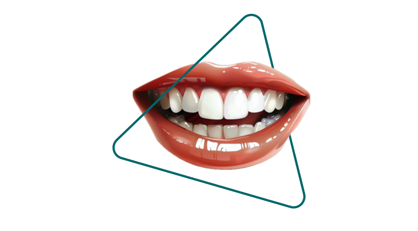
جراحی اصلاح طرح لبخند
لثه طبيعي در ناحيه طوق دندانها داراي فرم دندانه اي است كه به آن اسكالوپ ميگويند. ارتفاع و عرض اسكالوپ لثه يكي از مولفه هايي است كه در دندانهاي جلويي فكين به آن طرح لبخند يا smile design گفته ميشود. بطور خلاصه نسبت اندازه ي دندانهاي پيش و نيش و آسيابهاي كوچك بر اساس اصول زيبايي تعريف شده است. اين نسبتها در جوامع و نژادهاي مختلف و نيز بر اساس چهره ي هر فرد و ارتفاع و حجم لبها و مقدار ديده شدن لثه در طي يك لبخند طبيعي، متفاوت ميباشد. همانطور كه اشاره شد عوامل ديگري هم در يك لبخند زيبا موثر هستند براي مثال رنگ و درخشندگي دندانها و اندازه مناسب قوس فكي و ميزان اوربايت و اورجت دندانها را ميتوان ذكر كرد.به اين ترتيب جراحي لثه براي فرم دهي اسكالوب لثه اي و طراحي سايز مناسب دندانها قسمتي از طرح درمان براي ايجاد و بازسازي لبخندي زيبا و اصولي است. متعاقب آن ممكن است دندانها نياز به سفيد كردن يا بليچينگ داشته باشند و يا ترميمهايي از جنس كامپوزيت، روكشهاي زيركونيا، لمينيت هاي دنداني، و از اين دست براي بيمار انجام شود.اين درمان يك همكاري نزديك بين متخصصين رشته هاي پريو و ترميمي-زيبايي و گاهي متخصص ارتدنسي را مي طلبد .
Smile Design Corrective Surgery
The natural gingiva around the necks of the teeth has a scalloped shape, known as the gingival scallop. The height and width of this scallop are key factors in the aesthetics of the front teeth, a concept referred to as smile design. In simple terms, the proportional size of the incisors, canines, and premolars is defined according to aesthetic principles. These proportions vary among different populations and ethnicities and are also influenced by individuals’ facial features, lip height and volume, and the extent of gingival visibility during a natural smile.
As mentioned, other factors contribute to a beautiful smile, including the color and brightness of the teeth, the proper shape of the dental arch, and the degree of overbite and overjet. Therefore, gingival surgery aimed at reshaping the gingival scallop and designing the appropriate tooth size is an essential part of treatment to create and restore an attractive and harmonious smile.
Following this procedure, additional treatments may be required, such as teeth whitening (bleaching) or restorations using composite materials, zirconia crowns, dental veneers, and similar aesthetic solutions. This type of treatment requires close collaboration between periodontists, restorative and cosmetic specialists, and sometimes orthodontists.
جراحی لیفت لثه برای اصلاح لبخند لثهای
طبق تعريف طي يك لبخند طبيعي بايد چيزي حدود ٢ تا ٣ الي ٤ ميليمتر از لثه نمايان شود. ديده شدن مختصري لثه بويژه در خانمها لبخند را زيباتر ميكند در حالي كه در اآقايان ديده نشدن لثه ها ميتواند جذاب تر باشد. زماني كه لثه بيش از اين ميزان ديده شود اصطلاح لبخند لثه اي يا gummy smile براي آن بكار ميرود كه يك مشكل چند عاملي يا multifactorialبشمار مي آيد. لبخند لثه اي بر اساس ميزان ديده شدن لثه به سه دسته خفيف، متوسط، و شديد( بيش از ٨ ميليمتر ديده شدن لثه) تقسسم ميشود. بطور خلاصه علل آن عبارنتد از:
Delayed passive eruption
Gingival overgrowth
Skeletal abnormality; vertical maxillary excess
Overgrowth of pre-maxilla
Dental arch asymmetry and occlusal plane discrepancy
درمان اين مشكل بر اساس علت آن طراحي و اجرا ميشود. گاهي تلفيقي از عوامل دنداني، لثه اي، اسكلتي در ايجاد لبخند لثه اي دخالت دارند. به همين دليل درمان ممكن است نياز به همكاري متخصصين چندين رشته از جمله پريودنتيست، متخصص ارتدنسي، و جراح فك و صورت داشته باشد. جراحي ليفت لثه و حذف لثه ي اضافي و گاهي استخوان از اطراف طوق دندانها روشي است كه در درمان gummy smile با منشا لثه اي بكار ميرود. با انجام اين جراحي، تاج دندانها درشت تر و مقدار لثه كمتر. ديده خواهد شد .
Periodontal Surgery for Correction of Gummy Smile
According to the definition, in a natural smile, approximately 2-4 millimeters of gingival tissue should be visible. A slight display of gingiva, especially in women, enhances the beauty of the smile, whereas in men, minimal gingival exposure may be more attractive. When more than usual gingival tissue is visible, it is termed a gummy smile—a multifactorial problem. Gummy smile is classified as mild, moderate, or severe( more than 8 mm of gingival visibility). In summary, its causes include:
• Delayed passive eruption
• Gingival overgrowth
• Skeletal abnormality; vertical maxillary excess
• Overgrowth of the pre-maxilla
• Dental arch asymmetry and occlusal plane discrepancy
Treatment of this problem is based on identifying and addressing its underlying causes. Sometimes, a combination of dental, gingival, and skeletal factors contributes to the gummy smile. Therefore, treatment may require cooperation among specialists in several fields, including periodontist, orthodontist, and maxillofacial surgeon. Gingival lift surgery—which involves the removal of excess soft tissue, and occasionally bone around the necks of the teeth—is a method used for treating a gummy smile of gingival origin. This surgery results in the appearance of larger dental crowns and reduces gingival visibility and exposure .
جراحی افزایش طول تاج
جراحي افزايش طول تاج دندانها (CL), بطور كلي با هدف بهبود زيبايي (استتيك)، يا عملكرد (فانكشن) دندانها و يا هر دو انجام ميگيرد.
در جراحي CL استتيك، لثه و استخوان اطراف دندانهايي كه در محدوده ي ديد يا esthetic zone قرار دارند (كه عمدتا شامل ١٠ دندان در هر فك ميشوند )، با عمل جراحي افزايش طول تاج در موقعيت جديدي قرار داده ميشوند. محل قرار گيري جديد لبه لثه بر اساس مقادير از پيش تعيين شده و محاسبه شده ي طول و عرض دندانها نسبت به يكديگر ميباشد. اين جراحي نياز به همكاري نزديك متخصص جراحي لثه و متخصص ترميمي- زيبايي دارد تا ظاهر دندانها زيباتر و متناسب تر ديده شوند. چنانچه رنگ يا فرم كلي دندانها نياز به اصلاح داشته باشند يا دندانها دچار پوسيدگي يا سايش شده باشند معمولا بعد از جراحي CL، درمان هاي روكش دنداني، ونير كامپوزيت، يا لمينيت انجام ميشود.
جراحي CL فانكشنال نيز در مواردي انجام ميشود كه پوسيدگي يا ترك تاج دندان به زير لبه ي لثه گسترش پيدا كرده باشد و دندان نياز به ترميم يا روكش داشته باشد. در چنين شرايطي با انجام جراحي CL مقدار معيني از بافت لثه و استخوان از اطراف ناحيه ي پوسيدگي يا ترك برداشته ميشود. به اين ترتيب از قرار گرفتن لبه ترميم يا روكش در داخل بافت لثه يعني ناحيه اي كه محل قرار گيري الياف سوپراكرستال است ممانعت شده و استخوان زيرين دچار آسيب ديدگي و تحليل نميشود .
Crown Lengthening Surgery (CL)
Crown lengthening surgery (CL) is generally performed to enhance the aesthetics, function, or both of the teeth.
Aesthetic CL Surgery:
In aesthetic crown lengthening, the gingiva and surrounding bone of the teeth within the visible area (esthetic zone), typically including 10 teeth in each jaw, are repositioned surgically. The new position of the gingival margin is determined based on pre-calculated measurements of the proportional length and width of the teeth. This procedure requires close collaboration between a periodontist and a restorative/aesthetic specialist to achieve a more harmonious and attractive appearance of the teeth. If the color or shape of the teeth needs correction, or if the teeth are affected by caries or wear, additional treatments such as dental crowns, composite veneers, or laminates are usually performed after the CL surgery.
Functional CL Surgery:
Functional crown lengthening is performed when tooth decay or a fracture extends below the gingival margin, requiring restoration or a crown. In such cases, a specific amount of soft tissue and bone is removed around the affected area to prevent the restoration or crown margin from invading the attached supra-crestal fibers, which could damage the underlying bone.